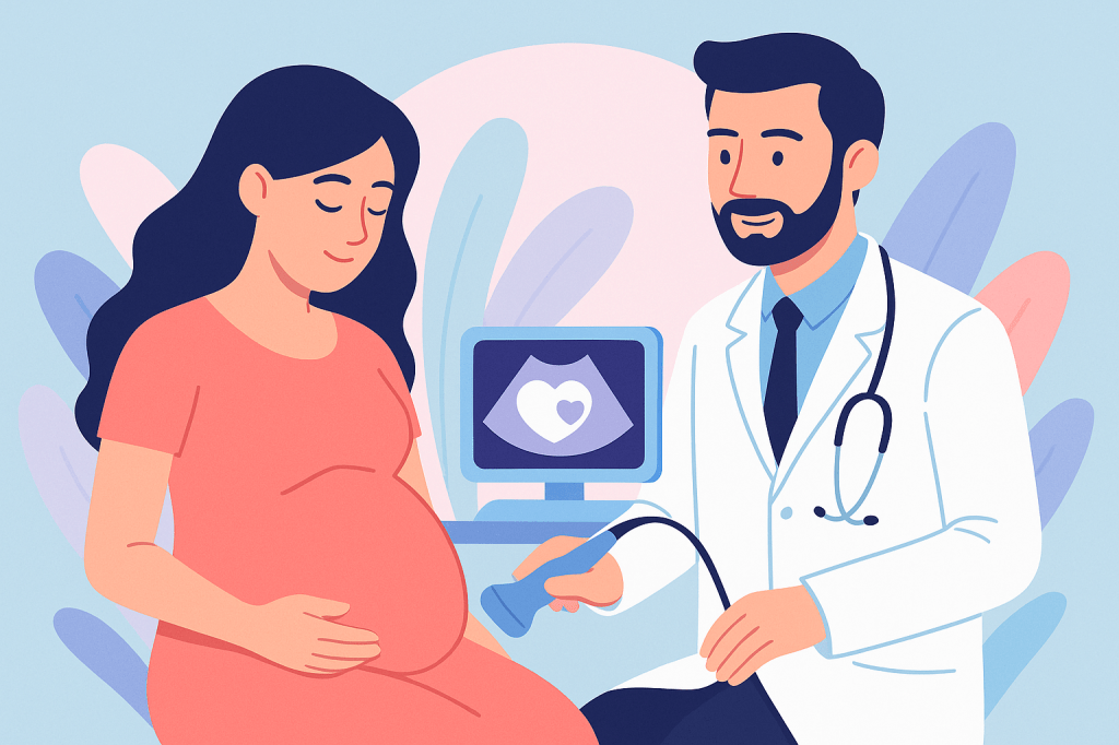
Fetal echocardiography is one of the best tests to study a baby’s heart during pregnancy. It gives valuable information that helps doctors plan care and, when needed, prepare for treatment after birth.
However, like all medical tests, it has some limitations. Knowing these helps set realistic expectations and reduces unnecessary worry.
1. The Test Is Very Good, But Not Perfect
- Most major heart problems can be seen.
- Very small or mild changes may not be visible.
- Some heart conditions develop later in pregnancy or after birth, even if the early scan looks normal.
2. Why Some Details May Be Hard To See
Factors that affect clarity are natural and nobody’s fault:
- Baby’s position during the scan
- Early pregnancy stage
- Less fluid around the baby
- Mother’s body build or previous abdominal surgery
3. Some Conditions Naturally Need Follow-Up
A few conditions are harder to diagnose fully before birth, such as:
- Very small holes (tiny ASD/VSD)
- Aortic coarctation
- Mild valve disease
- Abnormal lung vein drainage (TAPVC)
- In these cases, your doctor may suggest repeat scans.
4. Baby’s Heart Can Change With Time
The heart grows and develops throughout pregnancy.
Some conditions may improve, progress, or become clearer on later scans, so a repeat study is sometimes advised.
5. After-Birth Check Is Still Important
Even with a normal fetal echo, a routine newborn check is advised.
A postnatal echocardiogram may be recommended if there are clinical signs, symptoms, or risk factors.
6. Things to Keep in Mind
- The scan gives excellent guidance and reassurance.
- It helps doctors plan wisely and be prepared.
- A normal scan is very encouraging, but no test in medicine is 100% perfect.
7. How Accurate Is the Test?
Research studies have shown that fetal echocardiography can correctly identify many babies with significant heart problems. Overall sensitivity (ability to detect a problem when it is present) is reported around 60–80%, while specificity (ability to confirm a normal heart when no abnormality exists) is very high, around 98–100%.¹,²
In experienced centres and later gestational ages, the detection rate for major heart defects may be even higher.²
This means the test is highly reliable, especially when it shows a normal result, but it cannot guarantee 100% detection.
8. Conclusion
Fetal echocardiography is a highly reliable and valuable test, but results are best combined with clinical follow-up and good communication.
References (Where This Information Comes From)
1. Meta-analysis on diagnostic accuracy of fetal echocardiography (pooled sensitivity and specificity): Wu C, Chen B, Wang Q, et al. Diagnostic value of fetal echocardiography for congenital heart disease: A systematic review and meta-analysis. Medicine (Baltimore). 2015;94(43):e1759. DOI: 10.1097/MD.0000000000001759
2. High accuracy reported from specialist fetal cardiac centres and later gestation studies: DeVore GR, Polanco B, Sklansky M, Platt LD. The value of a cardiovascular profile score in the detection and management of fetal cardiac anomalies. Journal of the American Society of Echocardiography (JASE). 2024;37(6):677–690. (Includes high-detection performance reports in dedicated fetal cardiac units.)
